

Soft tissue injuries affect the muscles, ligaments, tendons, and other structures that support movement throughout the body. These injuries occur during accidents or everyday activities, and they can be difficult to assess through a physical examination alone. Magnetic resonance imaging, commonly known as MRI, offers a detailed view of these structures.
What Is an MRI?
An MRI is a diagnostic tool that produces detailed images of the inside of the body. Rather than using radiation, as X-rays and CT scans do, an MRI relies on a strong magnetic field and radio waves. These elements work together to generate signals from the water molecules present in the body’s tissues; a computer then translates those signals into clear, cross-sectional images.
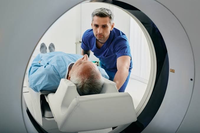
The equipment itself is a large machine with a cylindrical opening at its center. During the scan, a person lies on a movable table that slides into this opening. Different tissues can contain varying amounts of water, so the resulting images show contrast between structures such as muscle, cartilage, and bone.
What Is It Used For?
MRI is widely used to examine a range of injuries that involve the body’s soft structures. It produces high-resolution images, so it can help identify damage that is not visible through other methods. There are a variety of injuries that an MRI may identify, including:
- Ligament Tears: MRI can reveal tears in structures such as the anterior cruciate ligament (ACL) in the knee or the ligaments of the ankle.
- Tendon Injuries: An MRI may show inflammation, partial tears, or complete ruptures in areas like the Achilles tendon or the rotator cuff of the shoulder.
- Muscle Strains: Overstretched or torn muscle fibers can appear on an MRI, along with any associated swelling or bleeding within the tissue.
- Disc Problems in the Spine: The soft discs between the vertebrae may bulge or herniate, and an MRI provides a clear view of these structures and the surrounding nerves.
By showing the extent and location of an injury, an MRI helps support informed decisions about care and treatment planning.
What Should You Expect?
A person scheduled for an MRI can prepare by knowing the general steps involved. Before the scan, staff will ask about any metal implants, devices, or objects in the body, since the magnetic field interacts with metal. During the procedure, the individual lies still on the table as it moves into the scanner. The machine produces loud tapping and humming sounds while it captures images, and earplugs or headphones are usually provided. Remaining still is one of the main requests during the scan, as movement can blur the images. After the session, most people return to their normal activities right away.
Seek Professional Imaging Services
A soft tissue injury may call for a closer look, and professional imaging services provide the detailed information needed to guide the next steps toward recovery. Understanding how this imaging works, along with what it can reveal, allows a person to approach the process with clarity. A specialist will review the images afterward and interpret the results for you.
